Disc is a cushion like strecture lying between the vertebral bones. Jelly like substance within the disc(Nucleus pulposus) is responsible for the flexibility of back bone. Some times the jelly like substance comes out of its boundary and is called as disc prolapse .

Disc prolapse if occurs in the neck it causes neck pain with arm pain. If it occurs in low back it causes lowback pain with leg pain.Pt might also have weakness and numbness of arm or leg depending upon the severity of nerve compression.
Xray is the first investigation needed. MRI may be needed if pain or weakness is severe.
Mostly pain will subside with physiotherapy , rest and pain medications. Discectomy surgery might be needed if pt has persistent arm or leg .
Disc is a cushion like strecture which acts like a shock absorber and is needed for flexibility of back bone.When the disc gets dehydrated (loss of flexibility) because of wear and tear/ heavy mechanical work it is called as a degenerated disc. It commonly occurs in lumbar spine (Low back region)
Many pts with degenerative disc disease will be asymptomatic while some may have low back pain on bending and other strenous activities.
Xray is the first investigation and MRI might be needed if pain is severe.
The treatment is mainly physiotherapy and back strengthening exercises.
Age related wear and tear occuring in the back bones is called as spondylosis.
Lumbar spondylosis- occurs in the low back region
Cervical spondylosis – occurs in the neck region
Majority of the patients will be asymptomatic. Some patients will have neck pain in cervical spondylosis and low back pain in lumbar spondylosis.
Xray of the concerned part will be helpful in diagnosis.
The treatment is mainly physiotherapy and back/neck strengthening exercises.
Physiotherapy includes – traction., IFT and wax bath.
Low back pain arising out of injury to the muscles or ligaments around the low back region (Lumbosacral region) is called as lumbosacral strain / sprain.
Arises out of sudden bending activity or lifting heavy weight.
Patients might have varying degree of low back pain related to activities.
Xray might be needed in some patients.
The treatment is mainly physiotherapy , rest for short duration and pain medications.
Physiotherapy includes – traction., IFT and wax bath.
Fracture in the vertebral body disturbing the continuity in the alignment of back bones is called as spine fracture. Commonly occurs due to road traffic accident and fall from height.
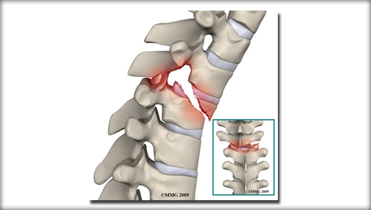
In simple fracture patient will have back or neck pain .In severe injury patient might have weakness of arms or legs.
Xray is the initial investigation . MRI and CT may be required dependin on the severity of the injury.
The treatment for simple fracture is rest and supportive appliances such as belt/ collar and orthosis. Complex fracture with displacement or with weakness might require surgery for stabilisation.
Infection occuring in the disc space is called as spondylodiscitis. Most commonly the infection is due to tuberculosis and rerely by other bacterias and fungus.
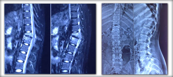
Patient will have pain , fever , loss of weight and loss of appetite. In severe case weakness can also develope.
Investigations needed are basic blood investigations( complete hemogram,CRP) X-ray and MRI. Biopsy ( testing the infected tissue) is needed to find out the organism causing the infection.
For initial stages of infection antibiotic medicines along with supportive appliances such as belt/ collar and orthosis is needed. In later cases with severe pain , deformity and weakness surgical intervention is needed. In case of tuberculosis infection 9 months of drug intake is needed.
The back bones are very flexible because of spring like disc substance and facet joints inbetween the vertebral bones . These spring like tissues becomes rigid and loses its flexibility in ankylosing spondylitis. It also affects other areas such as sacroiliac joints , hip joints and rib cages.
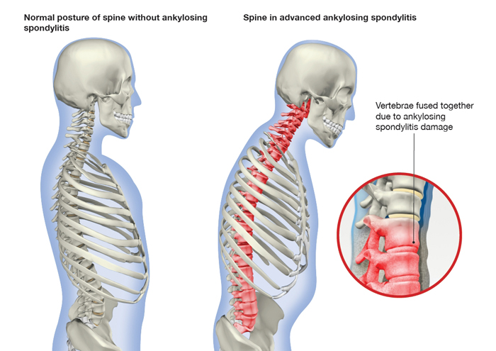
Pateints initially presents with lowback pain. In later stages patient might develop spinal deformity, loss of spinal movements and hip arthritis.
Investigation include blood investigations (complete hemogram, CRP and HLA B27), Xray of the involved region and MRI if needed.
Treatment includes supportive measures like pain medications , back exercises and deep breathing exercises. Rheumatological consultation is needed and newer drugs like disease modifying agents might be needed.
The spine is made of series of connected bones called as vertebrae. Spondylolisthesis is abnormal forward displacement of one vertebra over the other which occurs mainly in low back region. Spondylolisthesis occuring in young age may be due to abnormal development ( Dysplastic spondylolisthesis) or due to fractute in the connecting bone( Lytic spondylolisthesis). In old age degeneration is the main cause of spondylolisthesis(Degenerative spondylolisthesis).
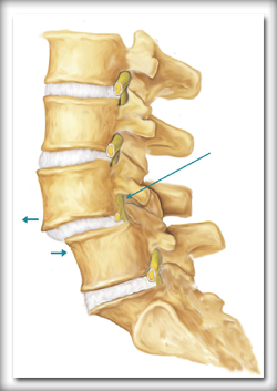
Patient usually presents with low back pain related to activities . It may be associated with leg pain if there is nerve compression.
Xray is the first line of investigation. MRI might be needed if the pain is persisting or if nerve compression is suspected.
Most of the patients settles down with physiotherapy- mainly back exercises , pain medications and activity modification. If the pain is severe and persisting surgical intervention may be indicated. Commonly done surgery is Lumbar decompression and pedicle screw fixation.
Any alteration in the normal shape of the spinal column is called as spinal deformity. If it is abnormal lateral bending of the spine it is called as scoliosis. If it is abnormal forward bending of spine it is called as kyphosis(hunch back). The deformity might arise out of birth defects which is called as congenital scoliosis or congenital kyphosis. Deformities arising during the adolescent years with out and birth defects is called as developmental or idiopathic deformity. In old age, degenerative changes can lead to deformity which is called as degenerative scoliosis or senile kyphosis.
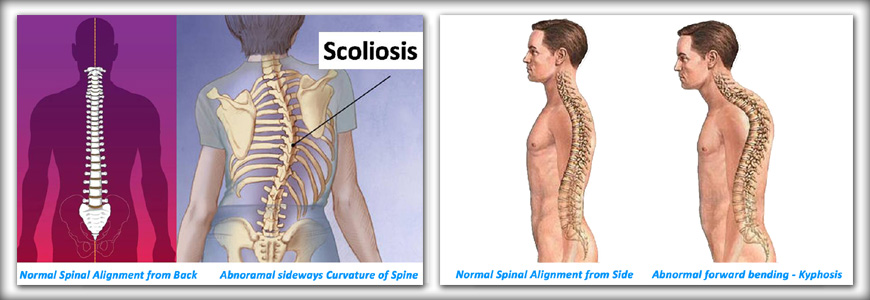
Patients might present with complaints of deformity in back, pain in back or sometimes weakness in limbs.
Xray is the initial investigation . MRI might be needed in some cases.
Treatment depends upon the severity of symptoms. In patients with mild deformity and minimal pain supportive appliances such as belt and orthosis is used along with back exercises. In severe deformity with cosmetic problems , weakness of limbs and significant pain surgery is indicated. Spinal deformity correction surgery is a complex surgery which requires expertise and special infrastrecture.
Nerves supplying the limbs arise from the spinal cord and travel along the limbs to its specific end point. For upper limb, nerves arise from cervical spine(neck) and for lower limb nerves arise from lumbar spine (lower back). If there is any compression on the nerve root(starting point of the nerve) the pain arising would travel along the course of the nerve to the limbs. This type of pain is called as radicular pain.
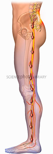
If the radicular pain is in the upper limb it is called as cervical radiculopathy and if the pain is in the lower limb it is called as lumbar radiculopathy. The most common cause of radiculopathy is disc prolapse.
MRI is the best modality to investigate the nerve root compression.
Treatment is based on the cause of radicular pain. In severe cases with persistent pain and weakness surgery to releive the nerve compression is needed.
Dysfunction of spinal cord arising out of compression is called as compressive myelopathy. Most commonly the compression is seen due to degenerative changes in cervical spine (neck) called as cervical myelopathy or in the thoracic spine (mid back) called as thoracic myelopathy.
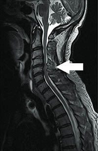
In cervical myelopathy both upper and lower limb is affected. In thoracic myelopathy only lower limb is affected.
Patient might present with clumpsiness of hands, difficulty in doing fine jobs with hands such as buttoning the shirt, change of handwriting and mixing the food. Change in gait pattern with unsteadiness and rigidity in limbs are other symptoms.
MRI is the best investigational modality for compressive myelopathy.
Treatment is mainly surgical decompression as early as possible. The disease will progress on delaying the surgery . The treatment is mainly to halt further progression.
Decreased bone strength is called as osteoporosis. Most commonly seen in old age postmenopausal women due to hormonal changes(lower levels of estrogen). Osteoporosis can also occur due to kidney diseases, alcoholism, surgical removal of ovaries and long term steroid intake.
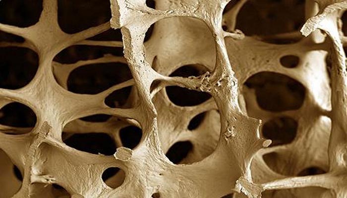
Patients with osteoporosis are prone to get fracture easily even with trivial fall. Most common regions to get fractured are back bone , wrist and hip bone. Apart from fracture patient may also have bone pain .
Xray is the basic investigational modality and DEXA scan is more specific for diagnosing osteoporosis.
Treatment is mainly medications. Calcium supplements with vitamin D is the main stay of treatment. Newer drugs like bisphosphonates and teriparatide injections aid in faster building up of bone strength.
The spine is made of series of connected bones called as vertebrae. The spinal canal is the central part of vertebrae through which the spinal cord travels. The lumbar canal is the central part of the lumbar vertebrae(Low back region). Narrowing of the lumbar canal due to degenerative changes leading to the compression of the nerves is called as lumbar canal stenosis.
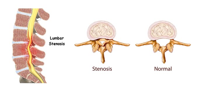
Nerves to the lower limbs arise from the lumbar region . In lumbar canal stenosis patients will have lowback pain associated with predominant leg pain . Leg pain occurs mainly while standind and walking and is called as claudication pain. Leg pain may be associated with numbness also.
Xray and MRI are the primary mode of investigation.
Physiotherapy in the form of back exercises and medications are the initial line of management. If the symptoms are persisting then surgical decompression of lumbar canal is needed. The surgical procedure is called as laminectomy .
Coccyx is the end part of back bone otherwise called as tail bone. Coccydynia is pain in the tail bone which occurs mainly during sitting position.
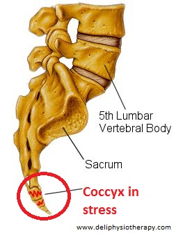
There will be history of trivial trauma – falling down in sitting position. Patient mainly complaints of pain over the tail bone on sitting over any hard surface , riding a two wheeler and pain worsens on getting up from sitting position.
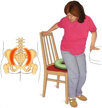
Usually no investigation is required. If pain is persistent xray / MRI may be suggested.
Sitting on a ring pillow is the main stay of treatment which avoids pressure over the tail bone. Supportive measures like pain medications and manipulation might help. In severe cases local steroid injection might help. Surgical removal of tail bone is done in extreme cases of pain.
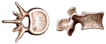
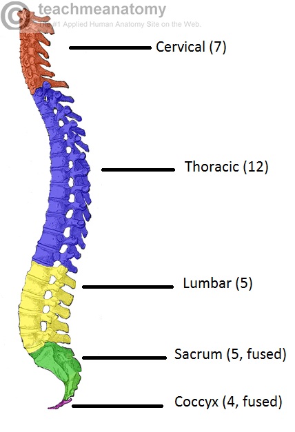
Vertebra is the basic bone in spinal column. It serves as building blocks of vertebral column. There are 33 vertebrae in our body.
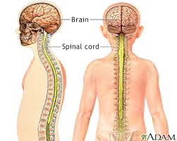
It is bundle of nervous tissue connecting brain with other parts of the body. It lies within the vertebral column.
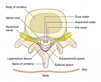
Originates from spinal cord and transmits signals to the muscles.
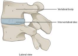
Cushion like substance between the vertebrae serving as shock absorbing system.
Upper part of vertebral column which constitutes neck.
Mid part of the vertebral column which extends from lower part of neck to mid back region.
Lower part of vertebral column which constitutes waist region.
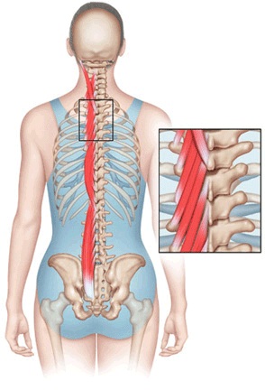
Muscles surrounding the vertebral column from neck to lower back. It gives stability to the vertebral column.
Weakness of both lower limbs due to injury to spinal cord. Complete weakness is called paraplegia and incomplete weakness is called praparesis.
– Weakness of both upper and both lower limbs . Complete weakness is called as Quadriplegia and incomplete weakness is called as Quadriparesis.
Bony discontinuity in vertebra / vertebral column.
A malignant tumour (cancer) spread to spinal column from else where.
Walking pattern of a person.
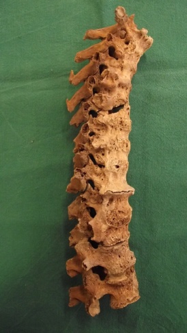
Abnormal bone formation along the sides of the vertebrae leading to stiffness.
Abnormal bone formation in the ligaments inside the spinal canal leading to spinal cord damage in few patients.
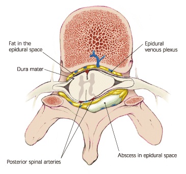
Pus formation inside the spinal canal surrounding the spinal cord.
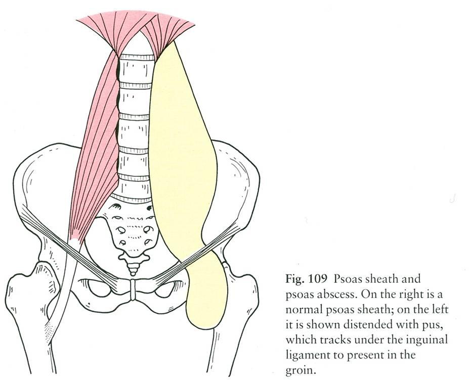
Pus formation or pus collection in the psoas muscle situated adjacent to the spinal column.
Weakness of ankle wherein a person is not able to walk on his heels.
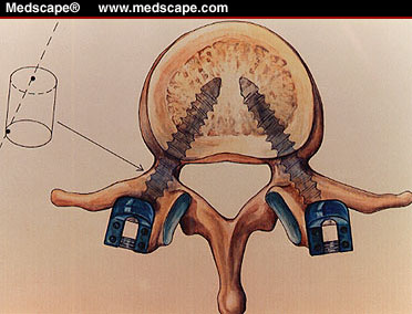
Implant inserted in to the pedicle of vertebra. It holds the vertebral body strongly. Used for spinal stabilization surgeries.
Exercises to strengthen the para spinal muscles which supports the spinal column.
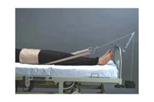
Weight applied to the pelvic/low back region . Mainly used for relief of low back pain by traction mechanism.
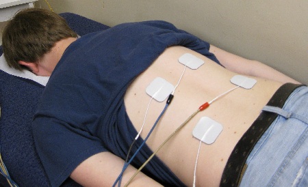
Small quantity of current is passed through the skin to the deeper tissues which helps to relieve the pain and is believed to stimulate healing process.
Biopsy is a minor surgical procedure where in a small quantity of the diseased tissue is taken for pathological examination under microscope. This is done to find out the exact nature of disease.
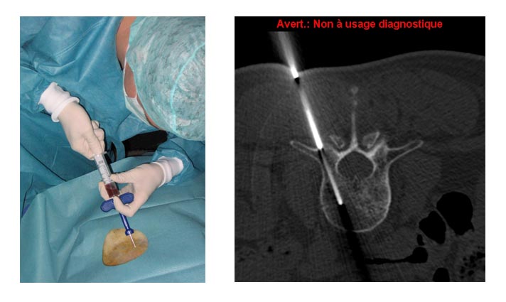
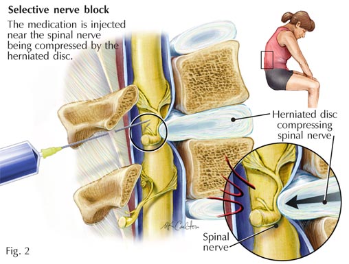
Pain in the arm or leg due to compression of nerve can be relieved by anesthetizing the nerve . Procedure of injecting the medicines to anasthetise thenerve is called as nerve root block.
When ever the urinary bladder is not functioning properly and not able to void urine we can insert a tube through the urinary meatus to drain the urine.
When the tube is inserted and kept inside it is called as indwelling urinary catheter. When the patient himself inserts the tube to drain the urine periodically it is called as self intermittent catheterization.

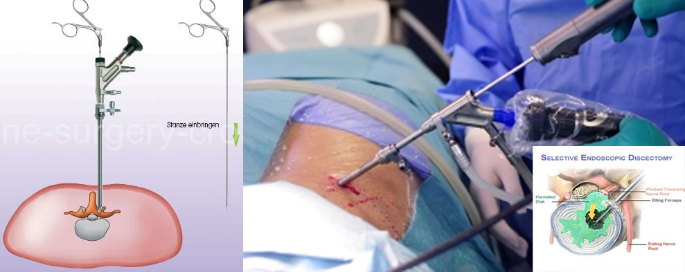
Spine surgery done through tubes (scopes) inserted through the skin . Minimises the muscle damage and post operative pain. Mainly used for treating lumbar disc prolapse.
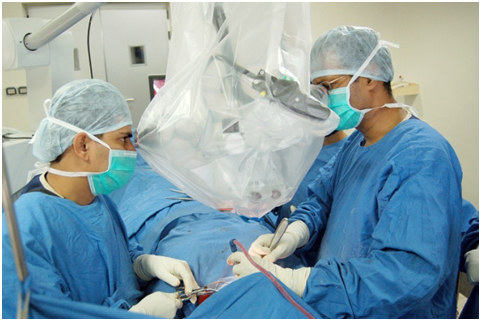
Spine surgery done using microscope . Microscope gives a good magnification and illumination and aids in safer spine surgery.
© 2016 - All Rights Reserved | Design and Developed By Rising Eminenze Technologies